DRSplus
True color confocal fundus scanner
NOW WITH IR AND AI SUPPPORT
Click to enlarge images


Choroidal Nevus
Compare IR and color image side by side to get a simultaneous view of retina and choridea.
NEW SOFTWARE 3.0.1 Sept 02, 2024
Clinical Benefits
- Swedish User Interface
- IR Images (gives choroidal view)
- Manual mode and fully auto
- AI (Artificial Intelligence) optional decission support; Diabetes (Glaucoma, AMD in late Q4-22)
- Very suitable for opticians
- 2.5 m.m. pupil, no need for dilation mostly
- Very simple to use, compact design
- Attractive pricing, very cost effective
- Only 11Kg! Makes it suitable for mobile scanning
- Tailored shipping case available
- Fully automatic with scanning protocols, saves time
- auto-alignment, auto-focus, auto-exposure, auto-capture
- Very high quality images, TRUE COLOR
- up to 80 degrees w. auto montage (from front of the eye).
- infrared LED (825-870 nm), white LED (440-650 nm), blue LED (440-475 nm)
- 10 Mpixel resolution
- Color
- Red Free
- Stereo imaging possible for 3D images on both Macula and ONH
- Even modest cataract works thanks to IR
- CE- marked
- Remote Exam feature (perform exam on a safe distance, from your web browser with full control, even from another location. Now even more useful due to Covid-19)
- Fully DICOM compatible (optional) for standardised integrations w. imaging systems and EMR
- DMWL (Worklist)
- C-STORE
Exclusive Features
- Remote Exam feature (perform exam on a safe distance, from your web browser with full control, even from another location. Now even more useful due to Covid-19)
- Remote service assistance
- Web API
- True white light, currently the only true color confocal scanner on the market
- Competitive pricing
- Full remote control in a web browser
- 3D on both Macula and ONH
Introducing DRSplus – a game changer in fundus imaging
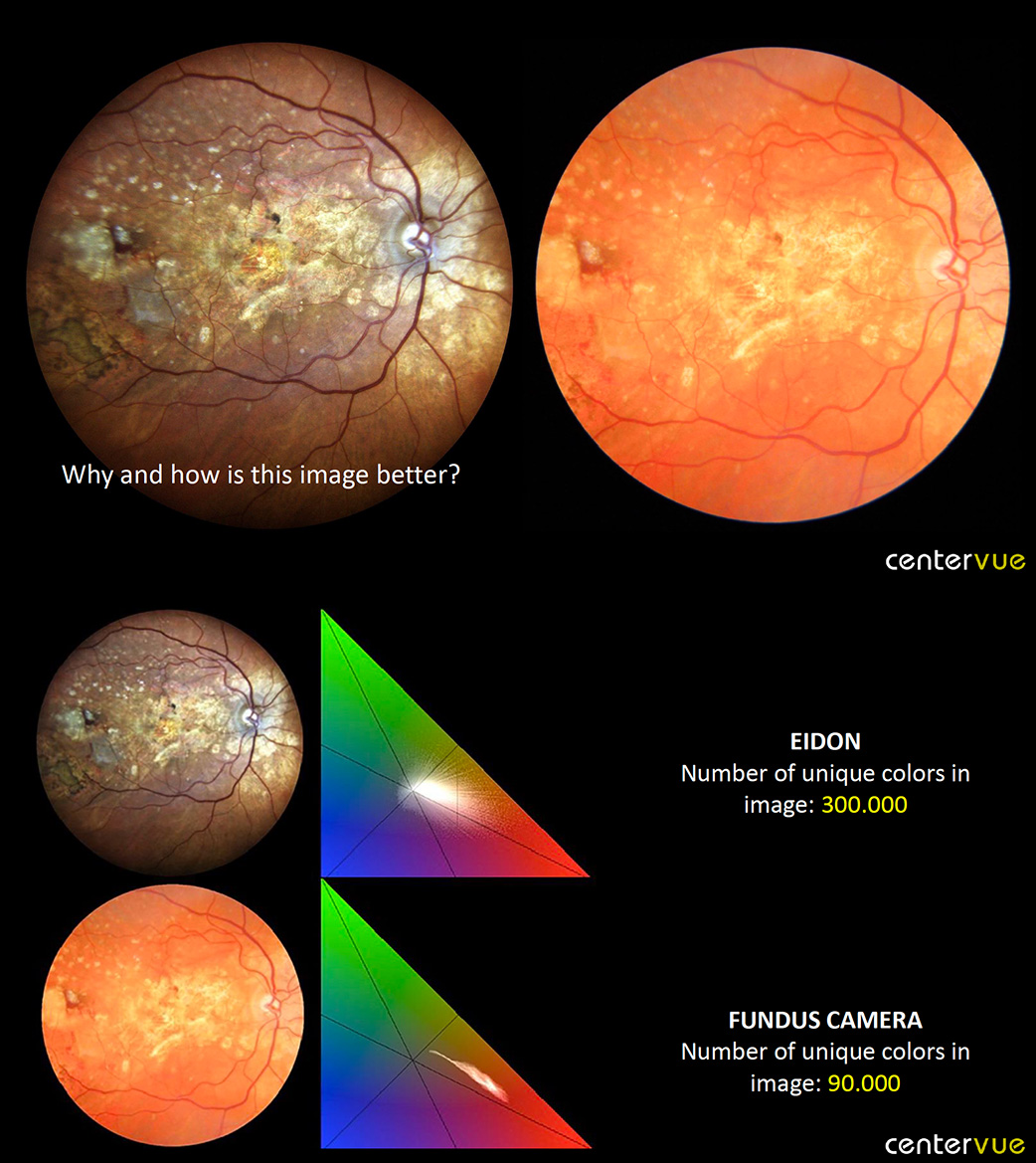
DRSplus confocal fundus imaging system by CENTERVUE uses white LED illumination to produce TrueColor and detail-rich images, setting a new effective and efficient standard of Retinal Imaging.
Confocal imaging is well known in ophthalmology as standard of high image quality. The main characteristic of confocality is to reduce or suppress any scattered or reflected light outside the focal plane. Thanks to this feature, DRSplus guarantees increased image sharpness, better optical resolution and greater contrast when compared to traditional fundus camera imaging. More over, the system uses a white light LED source, which includes the entire visible spectrum, to illuminate the retina and to capture TrueColor fundus images characterized by colours close to reality.


Stereo of disc and macula is possible.
Different Imaging Modalities
CENTERVUE DRSplus, in addition to TrueColor images, provides Red-Free, Blue and Red images of the retina. The system is able to capture external eye images and stereo view of ONH as well. Live IR supports during the images acquisition.

Fast and Fully Automated Operations
Fast Fully Automated operation requires minimal staff training to acquire the highest quality images, speeding up the examination time.
Fully-Auto capabilities, including Auto-Alignment, Auto-Focus, Auto-Exposure, Auto-Capture and Auto-Montage, make DRSplus the perfect Retinal Imaging System to fit needs of efficiency and time-saving. At the same time, the device is designed to optimize patient comfort during the exam, allowing for the improvement of workflows.
Remote Viewer
DRSplus Remote Viewer is a browser-based software license that allows for reviewing from any network computer on the same local area network (LAN), with password protection.
DRSplus offers embedded capabilities for network connectivity, for remote data review.
Remote Viewer enables dynamic viewing of DRSplus fundus images in your exam room or other remote location. In particular, it allows a detailed analysis of data in your office, facilitates patients’ education and enables data access on multiple review stations.

The minimum pupil size
DRSplus Confocal Technology permits imaging through pupils as small as 2.5 mm, speeding up the examination time while ensuring a comfortable patient experience.
DRSplus Non-myd capability makes fundus acquisition far more patient-friendly, by eliminating the need of dilating drops and dark environments. This also helps eliminating the wait for the pupil to dilate, with further benefits for the patient and saving time for the practitioner and office staff.

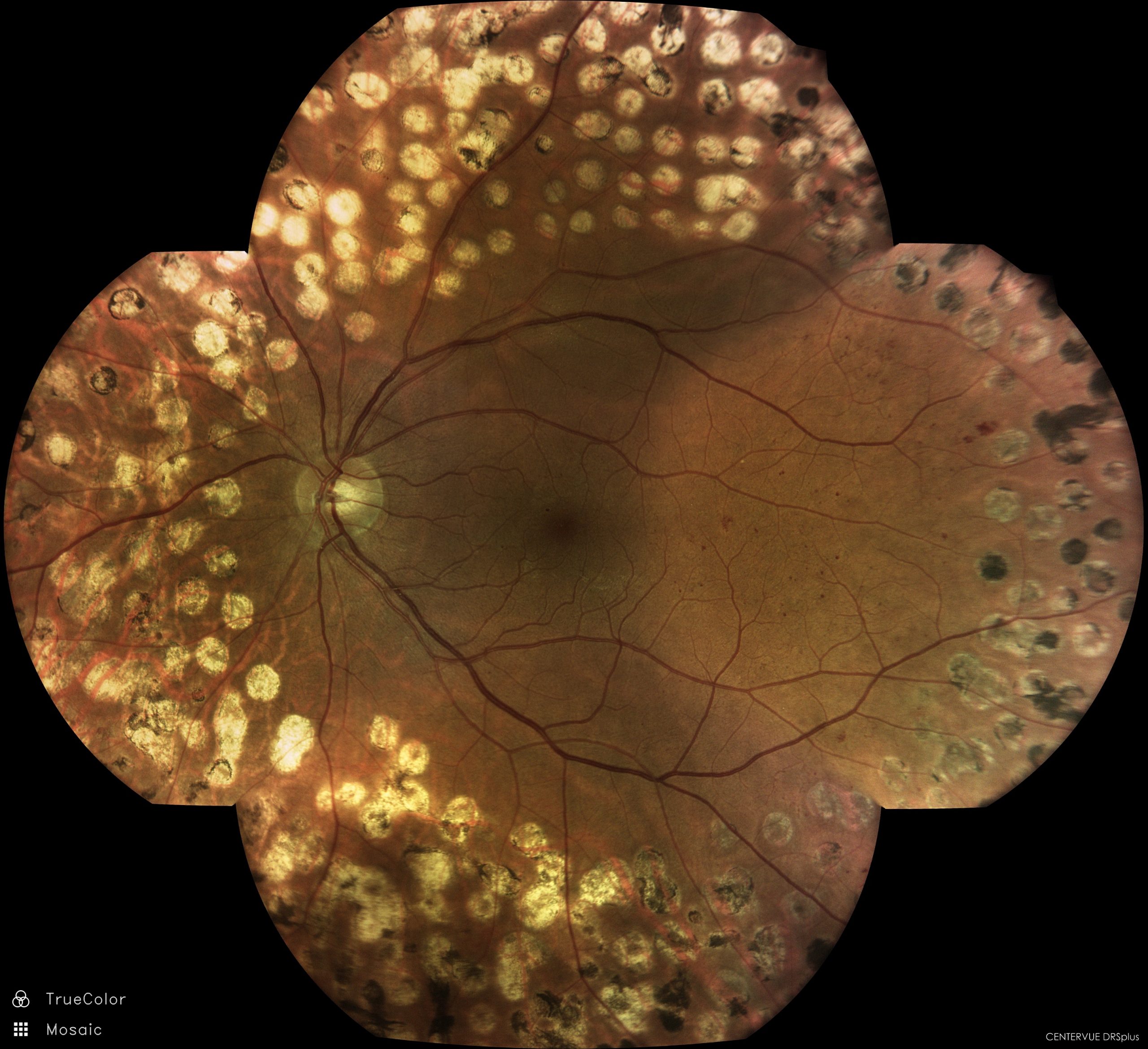
DRSplus sample image (click to enlarge)

The importance of true colors, WhatYouSeeIsWhatYouGet (WYSIWYG), for more detailed information, please see the White LED confocal vs. conventional Flash PDF above
The confocal true color technology differs from both traditional fundus cameras and Psedo color Confocal cameras, which is easy to see in the Image Gallery PDF.
Technical Specifications
Fundus Imaging System Features
- Field of ViewSingle image (W x H): 45° x 40°, Mosaic up to 9 fields (W x D): 83° x 78°
- Light SourcesWhite LED: 420-675 nm, Infrared LED: 825-870 nm
- Imaging ModalitiesTrueColor, Infrared, Red-Free*, Blue*, Red*, External Eye, Stereo**, Mosaic**
- Autofocus Adjustment Range-15D to +15 D
- Automatic OperationsAuto-alignment, Auto-focus, Auto-exposure, Auto-capture, Auto-montage
- Minimum Pupil SizeNon-myd 2.5 mm
- Working Distance25 mm
- Image Size10 Megapixels
- Resolution77 pixel/degree
- Fixation TargetsInternal/External**
- Dynamic Programmable Internal Fixation TargetCentral, Nasal, Temporal, Central-Nasal, Superior, Inferior, Superior-Temporal, Superior-Nasal, Inferior-Temporal, Inferior-Nasal
Computer
- DisplayIntegrated 10.1″ (1280 x 800) Color, Capacitive, Multi-touch
- Hard DriveSSD ≥ 480 GB
- InterfacesUSB port 2.0 x 3, Gigabit Ethernet Port x 1
- Export / Importjpeg, pdf, DICOM**, Web API**
- Remote Viewer**Up to a maximum of 5 remote stations
Dimensions/Power supply
- Size (W x H x D)300 mm (11.8”) x 450 mm (17.7”) x 650 mm (25.6”)
- Weight11 Kg (24 lbs)
- Rated Voltage100-240 VAC
- Frequency50-60Hz
- Power Consumption60W
- Electrical ClassIEC 60601-1 Class I
*Digital Filters
**Optional
The importance of true colors, WhatYouSeeIsWhatYouGet (WYSIWYG), for more detailed information, please see the White LED confocal vs. conventional Flash PDF above
The confocal true color technology differs from both traditional fundus cameras and Psedo color Confocal cameras, which is easy to see in the Image Gallery PDF.